Deeper clinical insights with revolutionary urinalysis
Understand your SediVue Dx results
A guide to urine sediment images and information for interpreting in-house results
Results reporting | Understanding bacteria | Automated sample messages | Reviewing images
Results reporting
Blood cells
White blood cells (WBCs)
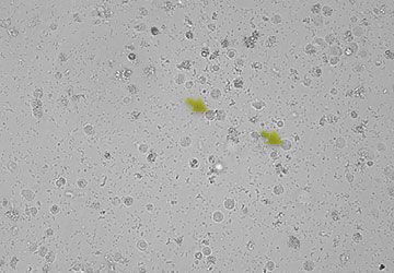
Image tag: WBC
| Result | |||
|---|---|---|---|
| None detected (The element has not been detected, or there are not enough recognizable features to classify.) |
<1/HPF (Some features were found in the sample; however, the quantity is below the clinical reporting threshold.) |
Quantitative numerical result/HPF | >50/HPF |
Red blood cells (RBCs)
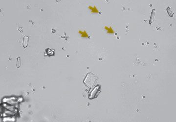
Image tag: RBC
| Result | |||
|---|---|---|---|
|
None detected |
<1/HPF (Some features were found in the sample; however, the quantity is below the clinical reporting threshold.) |
Quantitative numerical result/HPF | >50/HPF |
Bacteria
Rods
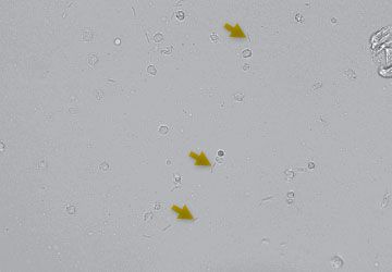
Image tag: N/A*
| Results | ||
|---|---|---|
| None detected (The element has not been detected, or there are not enough recognizable features to classify.) |
Suspect presence (Some recognizable features of an element (cocci, rods, casts) are present; however, the quantity and detail is insufficient to report as “present.”) |
Present (There is high confidence that bacteria are present in the sample.) |
Cocci
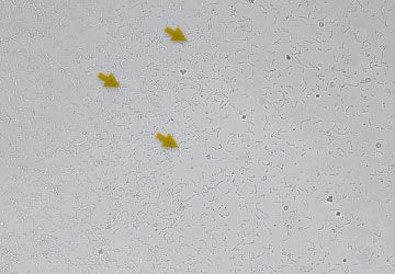
Image tag: N/A*
| Results | ||
|---|---|---|
| None detected (The element has not been detected, or there are not enough recognizable features to classify.) |
Suspect presence (Some recognizable features of an element (cocci, rods, casts) are present; however, the quantity and detail is insufficient to report as “present.”) |
Present (There is high confidence that bacteria are present in the sample.) |
*To avoid blocking your visual interpretation, the analyzer classifies and counts all bacteria without tagging them.
NOTE: Bacteria results may be confounded by other debris and artifacts in the sample (e.g., sperm, crystalline debris).
Epithelial cells
Squamous
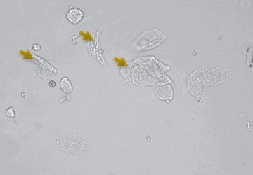
Image tag: sqEPI
| Results | |||||
|---|---|---|---|---|---|
| None detected (The element has not been detected, or there are not enough recognizable features to classify.) |
<1/HPF (Some rare features have been found in the sample; however, the quantity is below the clinical reporting threshold.) |
1–2/HPF | 3–5/HPF | 6–10 /HPF | >10/HPF |
Nonsquamous
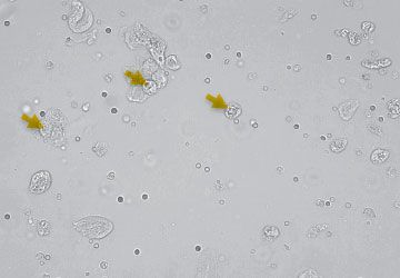
Image tag: nsEPI
| Results | |||||
|---|---|---|---|---|---|
| None detected (The element has not been detected, or there are not enough recognizable features to classify.) |
<1/HPF (Some rare features have been found in the sample; however, the quantity is below the clinical reporting threshold.) |
1–2/HPF | 3–5/HPF | 6–10 /HPF | >10/HPF |
Casts
Hyaline
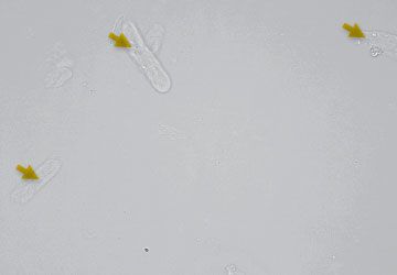
Image tag: HYA
| Results | ||
|---|---|---|
| None detected (The element has not been detected, or there are not enough recognizable features to classify.) |
Suspect presence (Some features of casts are present; however, there may not be enough to provide a >1/LPF result. Image review is strongly recommended for confirmation.) |
>1/LPF |
Nonhyaline (e.g., granular, waxy)
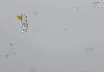
Image tag: nhCST
| Results | ||
|---|---|---|
| None detected (The element has not been detected, or there are not enough recognizable features to classify.) |
Suspect presence (Some features of casts are present; however, there may not be enough to provide a >1/LPF result. Image review is strongly recommended for confirmation.) |
>1/LPF |
Crystals
Unclassified (all other crystals)
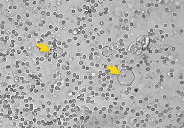
Image tag: CRY
| Results | |||||
|---|---|---|---|---|---|
| None detected (The element has not been detected, or there are not enough recognizable features to classify.) |
<1/HPF (Some rare features have been found in the sample; however, the quantity is below the clinical reporting threshold.) |
1–5/HPF | 6–20 /HPF | 21–50 /HPF | >50/HPF |
Calcium oxalate dihydrate
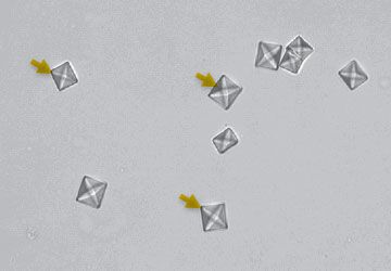
Image tag: CaOxDi
| Results | |||||
|---|---|---|---|---|---|
| None detected (The element has not been detected, or there are not enough recognizable features to classify.) |
<1/HPF (Some rare features have been found in the sample; however, the quantity is below the clinical reporting threshold.) |
1–5/HPF | 6–20 /HPF | 21–50 /HPF | >50/HPF |
Struvites
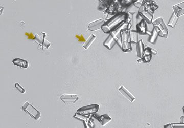
Image tag: STR
| Results | |||||
|---|---|---|---|---|---|
| None detected (The element has not been detected, or there are not enough recognizable features to classify.) |
<1/HPF (Some rare features have been found in the sample; however, the quantity is below the clinical reporting threshold.) |
1–5/HPF | 6–20 /HPF | 21–50 /HPF | >50/HPF |
Ammonium biurate
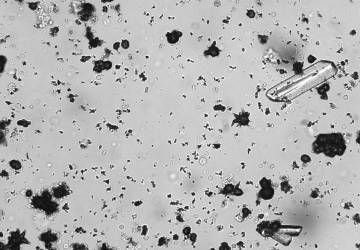
Image tag: AmmBi
| Results | |||||
|---|---|---|---|---|---|
| None detected (The element has not been detected, or there are not enough recognizable features to classify.) |
<1/HPF (Some rare features have been found in the sample; however, the quantity is below the clinical reporting threshold.) |
1–5/HPF | 6–20 /HPF | 21–50 /HPF | >50/HPF |
Bilirubin
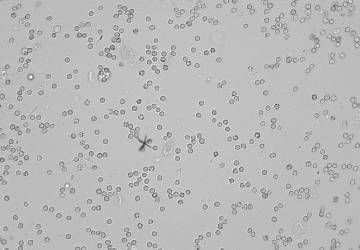
Image tag: BILI
| Results | |||||
|---|---|---|---|---|---|
| None detected (The element has not been detected, or there are not enough recognizable features to classify.) |
<1/HPF (Some rare features have been found in the sample; however, the quantity is below the clinical reporting threshold.) |
1–5/HPF | 6–20 /HPF | 21–50 /HPF | >50/HPF |
Understanding bacteria
Bacteria results
| Bacteria result (rods/cocci) | Definition | Possible reason for validation | Recommended next steps |
|---|---|---|---|
None detected |
The element has not been detected or there are not enough recognizable features to classify. | Patient has clinical signs or a history of persistent UTIs. | If visual review of images is negative and patient has no clinical signs or history, bacteriuria is unlikely. No further action is necessary. |
Suspect presence |
Some recognizable features of an element (cocci, rods, casts) are present; however, the quantity and detail is insufficient to report as “present.” | Crystalline or amorphous debris is common in canine and feline samples (especially free catch). |
Review visually: If visual review is conclusive:
If visual review is inconclusive:
|
Present |
There is a high confidence that bacteria are present in the sample. | Bacteria results may be confounded by other debris and artifacts in the sample (e.g., sperm, crystalline debris). |
If visual review of images is confirmatory and/or the patient has clinical signs or history, bacteriuria is likely. Diagnose and manage based on your interpretation. If visual review is inconclusive:
|
Top 4 questions about bacteria results
A result that reads "suspect presence" means that the SediVue Dx analyzer has detected enough uniformly sized, small structures in the sample to deem the result positive. However, there is not enough evidence to differentiate bacteria from crystalline or amorphous debris, which are both common in canine and feline samples, especially those obtained via free catch.
Here’s what we recommend doing:
First, review the images to determine if bacteria can be visually identified. If so, trust your visual interpretation of the sample and move on to a diagnosis, treatment, or follow-up appointment. Make a note of your findings in the patient record, and unless you feel the need for culture or sensitivity testing, no further action is necessary.
If, after reviewing the images, you are still not sure, ask these questions:
- Are white blood cells (WBCs) or red blood cells (RBCs) present?
- Does your patient have clinical signs?
- Is there a recurring history of lower urinary tract issues?
If the answer to any of the questions is "yes," perform the SediVue Bacteria Confirmation Kit or a dry prep (air-dried, stained cytological slide) to conclusively confirm the presence of bacteria. Watch our short videos.
If the absence of bacteria can clearly be determined in the images and there are no other supporting clues (active urine sediment, patient history, etc.), then the presence of bacteria is unlikely. However, by performing a new run with the SediVue Bacteria Confirmation Kit, you can have the confidence you need in just 3 minutes.
Key takeaways
- "Suspect presence" = sample contains uniform, small structures
- Results could be bacteria, crystalline debris, or amorphous debris
- Review images for additional evidence
- Presence of clinical signs and/or recurring history of lower urinary tract issues warrant a dry prep
If the SediVue Dx analyzer reports that a specimen is positive for bacteria and the culture result from the laboratory says negative, it is possible that both are correct. Even when best practices are followed, discordance between microscopic evaluation and culture results may occur.
Results of "no growth" on culture when bacteria are seen in the urine sediment may occur for the following reasons:
- Bacteria may have been visualized microscopically but may be dead (nonviable), especially if the animal is currently on antibiotics or had previously or recently been treated with antibiotics at the time of sample collection. Other factors that may inhibit or prevent bacteria growth in culture include exposure of the sample to temperature extremes, extremes of urine pH (≤4 or ≥9), or inhibition by white blood cells (in urine with "too numerous to count" white blood cells).
- The "organisms" identified with microscopy could have been crystalline or amorphous debris in the urine that was misidentified as bacteria, called pseudobacteria (particularly with unstained urine sediment examination).
- Random motion of small colloidal particles, known as Brownian motion, can falsely appear to be cocci bacteria (particularly with unstained urine sediment examination).
- Rarely, anaerobic bacteria may be visualized on urinalysis but not grown in aerobic cultures.
- If the urine sample was stained in-clinic prior to manual microscopic examination, the stain may have been contaminated with bacteria. Stains should be changed regularly.
Positive results on culture when bacteria are not identified during the urine sediment analysis may occur for the following reasons:
Bacterial colony counts are too low to be visualized on sediment analysis, for example in very dilute urine, following incomplete or unsuccessful antibiotic therapy or in the case of localized pyelonephritis. In situations where the clinical history is suggestive of urinary tract infection or an active urine sediment is present, urine culture should be considered even in the absence of bacteriuria on urinalysis.
Key takeaways
- Possible reasons for discordant results include the following:
- Dead (nonviable) bacteria
- Conditions that inhibit growth (temperature, pH, WBCs)
- Misidentified crystalline or amorphous debris
- Sample contaminated during staining
- Motion of small colloidal particles (Brownian motion)
- Very dilute urine
- Suggest urine culture in patients with a history of urinary tract infection or active urine sediment
A dry prep (air-dried, stained cytological slide) can be considered an alternative to running the SediVue Bacteria Confirmation Kit if you cannot visually confirm the absence or presence of bacteria in the images but suspect bacteria presence based on other supporting clues (active urine sediment, patient with clinical signs, or a recurring history of lower urinary tract issues) that could lead you to a diagnosis. A dry prep is a quick, easy way to validate the absence or presence of bacteria and distinguish it from amorphous or crystalline debris, and it can be done in 3–5 minutes!
NOTE: On rare occasions, bacteria can be found in the sample when there are no other supporting clues or clinical signs.
Key takeaways
- Perform a dry prep when you cannot visually confirm the presence of bacteria AND any of the following are present:
- Active urine sediment
- Clinical signs
- Recurring history of lower urinary tract issues
- Watch a short video to learn how to perform a dry prep in 3–5 minutes.
Bacteria tags on the images could be overwhelming and block your visual interpretation. Rest assured that even though bacteria are not tagged, the SediVue Dx analyzer is still counting and classifying them.
Key takeaways
- Absence of tags improves visual review
- SediVue Dx analyzer quantifies and classifies bacteria
Urinalysis versus culture
Understanding possible discordant results
| Bacteria result (rods/cocci) | Culture result | Causes for discordant results |
|---|---|---|
Present |
No growth |
*Particularly with unstained urine sediment examinations. |
None detected |
Positive |
|
Automated sample messages
Automated sample messages
Your SediVue Dx Urine Sediment Analyzer may include sample messages in the patient report. These messages are generated based on numerical results and are intended to provide you with further insight and guide you with recommended next steps.
Analyzer message
Confirm with one of the following: image review; the SediVue Bacteria Confirmation Kit; air-dried, stained cytological preparation ("dry prep"); or urine culture.
Definition
Some recognizable features of an element (cocci, rods, casts) are present; however, the quantity or details are insufficient to report as “present.” Debris commonly found in canine and feline urine can be confused with bacteria.
Guidance
Review the images. If images do not provide visual confirmation of bacteria or the sample is too crowded with cellular or crystalline debris, the next step would be to use the SediVue Bacteria Confirmation Kit. The Kit will dissolve the excess debris for better visualization in the images. If the Kit does not dissolve an adequate amount of material and the images still do not provide visual confirmation, but patient has clinical signs or there are other clues present (white blood cells, etc.), it is recommended to perform a dry prep to confirm.
If bacteria are obvious in the images and the patient has clinical signs, a dry prep is not necessary, but you can follow up with a culture and sensitivity (MIC) test if you want to further classify the bacteria as well as test for sensitivity to certain antibiotics.
Analyzer message
Images crowded. Review images and follow the guidance below to determine next steps.
Definition
When a urine sample is crowded and the edges of the elements overlap, the SediVue Dx convolutional neural network may have difficulty discerning the elements from one another.
Guidance
- When images provide clinical insight: No dilution needed, add comments to patient record.
- Moderate amount of cells or crystalline material: Dilute 1:5 with 0.9% normal saline and rerun.
- Marked amount of cells or crystalline material: Dilute 1:10 with 0.9% normal saline and rerun.
Note: You will only be charged for the first run from the same patient ID within a 24-hour period.
Analyzer message
Review images to confirm results.
Definition
The convolutional neural network cannot verify the quality of focus in the images.
Guidance
- The sample may have very little sediment (e.g., “normal”) or contain air bubbles, or the analyzer is dirty.
- If the expected results do not align with the image review, rerun the sample.
- If the message appears with several consecutive samples, the analyzer may require cleaning.
Note: You will only be charged for the first run from the same patient ID within a 24-hour period.
Crystals can come in a variety of different shapes, sizes, and presentations. Urine pH, specific gravity, sample preparation and handling, and drugs can all play a part in crystal formation. Crystals in small numbers (e.g., struvites) may be normal for some dogs, but others (e.g., cystine) may signify disease processes. The following Smart Flags are designed to provide further clinical insight into the presence of crystalluria.
Analyzer message
Crystalline debris detected.
Definition
Crystalline debris can be abundant and variable in size and presentation in some samples. Due to background density, the presence of large amounts of crystalline debris can affect the identification of other formed elements in the sample. This flag is displayed when crystalline debris has been detected by the algorithm. The neural network algorithm has been trained to exclude crystalline debris from the unclassified crystal (CRY) category.
Guidance
When this flag is present, users will be notified so they can be more discerning about the bacteria result, as very small particles of debris can resemble bacteria.
Analyzer message
Consider evaluation of urine protein:creatinine ratio.
Definition
When this message appears, the urine chemical results indicate the presence of protein. A UPC ratio can be used to quantify protein loss in the urine as it is unaffected by urine volume or concentration. It has been incorporated into the International Renal Interest Society (IRIS) Guidelines on Staging and Treatment of Chronic Kidney Disease (CKD) as an important monitoring tool at all stages.
Guidance
A UPC ratio should be performed after urinalysis with urine sediment examination. It is not recommended for use if there is an active urine sediment, as inflammatory conditions in the urinary tract will increase protein and negate the usefulness of the ratio.
Analyzer message
Recommend reevaluating proteinuria after resolution of active sediment.
Definition
When this message appears, the urine chemical results indicate the presence of protein in addition to an active urine sediment (red blood cells [RBCs] and/or white blood cells [WBCs] and possibly bacteria).
Guidance
First, resolve the infection. Once the urine sediment becomes inactive, consider running a UPC ratio to quantify protein loss.
For a complete list of automated sample messages, consult the SediVue Dx Urine Sediment Analyzer Operator's Guide.
Reviewing images
Guidance for reviewing images
Reviewing images will validate the numerical data provided and supplement the SediVue Dx analysis.
- Tap on an image to make it full screen.
- Reverse the contrast to see details such as cell nuclei.
- Zoom in up to 200% to see smaller elements.
- Turn image tags on or off.
- Select an image to add to the patient record or print an area of interest.
- Use the large arrows to scroll through images.
- Seventy high-resolution images are taken for every sample run.
- Images have been prioritized based on the absence or presence of formed elements and the clinical significance of each element found.
- Highest scoring images are displayed first.
- Select View all images to easily scroll through all 70 images.
- Classified elements may be tagged (labeled).
- Depending on the type of element, its concentration, and sample, you may not see tags.
- Whether a tag is present or not, all formed elements are classified by the analyzer and reflected in the result.
- For Cornerstone Software users, three prioritized images are automatically saved to the patient record and transmitted to VetConnect PLUS. You may select up to three additional images to save to the patient record and VetConnect PLUS.
- For all other practice management software, the first image will be included with the results PDF.
- Include comments about findings you consider noteworthy.
- From the patient results screen, tap Add comments. Your comments will be automatically uploaded to VetConnect PLUS and, if applicable, your practice management software.
Image tags are not available for “none to rare” for all elements as well as “suspect presence” for casts and all bacteria categories. However, all elements that are found are identified, classified, and counted. Tagging bacteria could be overwhelming and block your visual interpretation. The SediVue Dx Urine Sediment Analyzer classifies and counts all bacteria without tagging it.
A guide to all urine sediment images from the SediVue Dx analyzer
Reference bar = 20 microns
Blood cells
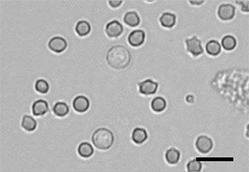
Figure 1. Red blood cells
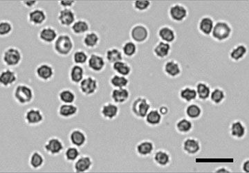
Figure 2. Crenated red blood cells
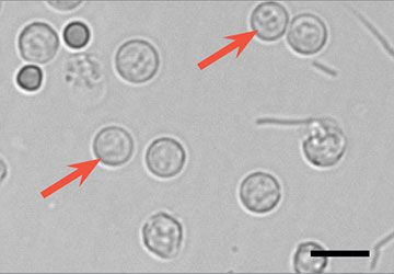
Figure 3. White blood cells
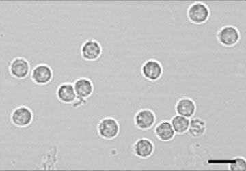
Figure 4. White blood cells
Epithelial cells
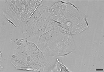
Figure 5. Squamous epithelial cells
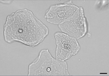
Figure 6. Squamous epithelial cells
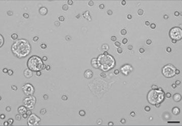
Figure 7. Numerous transitional (nonsquamous) epithelial cells with red and white blood cells
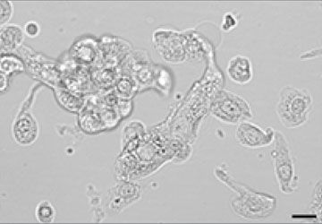
Figure 8. Numerous transitional (nonsquamous) epithelial cells (possible transitional cell carcinoma; confirm with dry prep)
Bacteria
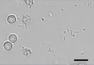
Figure 9. Rods with white blood cells
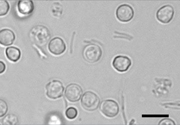
Figure 10. Rods with white and red blood cells
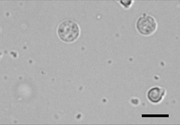
Figure 11. Cocci with white blood cells
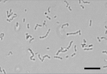
Figure 12. Cocci in chain
Casts
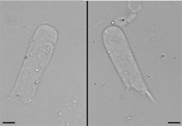
Figure 13. Left and right, hyaline cast
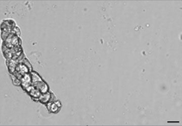
Figure 14. Cellular (nonhyaline) cast
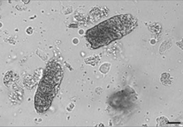
Figure 15. Numerous granular (nonhyaline) casts
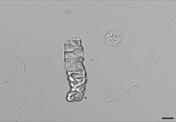
Figure 16. Waxy (nonhyaline) cast
Crystals
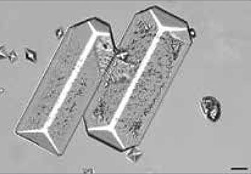
Figure 17. Large struvite crystals
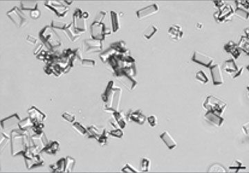
Figure 18. Numerous small struvite crystals
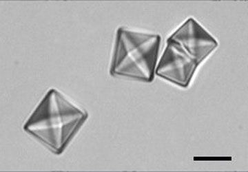
Figure 19. Large calcium oxalate dihydrate crystals
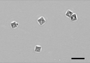
Figure 20. Numerous calcium oxalate dihydrate crystals
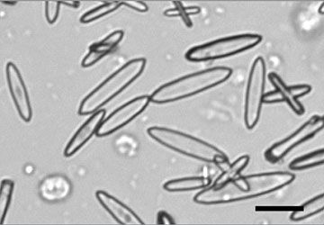
Figure 21. Calcium oxalate monohydrate (picket fence) crystals
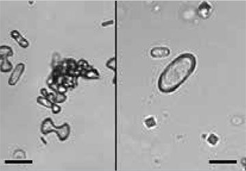
Figure 22. Calcium oxalate monohydrate crystals; left, dumbbells; right, hemp seed
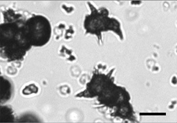
Figure 23. Ammonium biurate (thorn apple) crystals
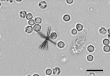
Figure 24. Bilirubin crystal with white blood cells
Figure 25. Cholesterol crystals
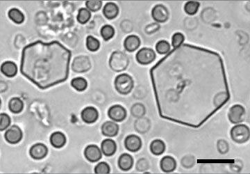
Figure 26. Cystine crystals with red blood cells
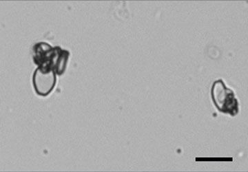
Figure 27. Uric acid crystals
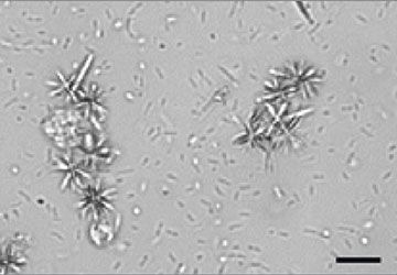
Figure 28. Likely drug-related crystals
Miscellaneous
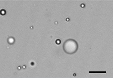
Figure 29. Lipids
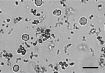
Figure 30. Amorphous crystalline debris
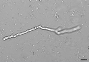
Figure 31. Hyphae
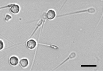
Figure 32. Sperm with white blood cells
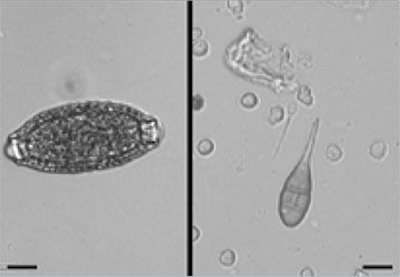
Figure 33. Left, Pearsonema spp. (Capillaria spp.) ova; right, macroconidia
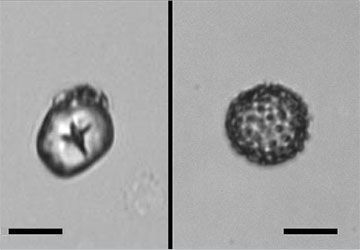
Figure 34. Left, glove powder; right, pollen
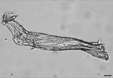
Figure 35. Fiber
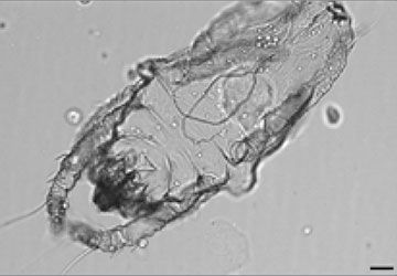
Figure 36. Dust mite
Get answers to common questions about results and images: FAQs
Which patients need a complete urinalysis?
Data supports the value of a complete urinalysis in "healthy" patients, those that are clinically ill, and those with conditions that require ongoing monitoring. See why.


Support
SediVue Dx support
Read FAQs and product how-to resources to help troubleshoot and get the most out of your SediVue Dx analyzer.
Contact us
Need more help? Get product answers and information or talk to Customer Support today.
Training courses
Take complimentary courses on SediVue Dx analyzer operation and urinalysis. Get video tutorials on daily protocols and procedures.