Article
Common pitfalls in veterinary imaging: how to avoid images that limit diagnostic confidence
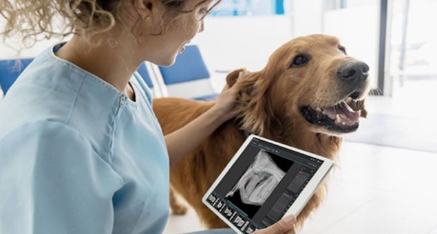
High-quality diagnostic images play a central role in clinical decision-making in veterinary medicine. Even with modern digital radiography systems, blurred, distorted, or nondiagnostic images can still arise, delaying diagnosis and contributing to retakes and additional radiation exposure.
Dr. Emma Turner, MSc, DVM
June 2, 2026
Key takeaways
- Most imaging challenges start at acquisition. Motion, positioning errors, technique selection, and incomplete studies are common, but largely preventable, causes of nondiagnostic images.
- Small adjustments can make a big difference. Thoughtful restraint, appropriate sedation, consistent positioning aids, and standardized exposure techniques help improve image clarity and reduce retakes.
- Complete, well-positioned studies support faster diagnosis. Capturing the correct views with full anatomic coverage the first time strengthens diagnostic confidence and workflow efficiency.
- Modern tools help teams image with confidence. Integrated DR systems, positioning guidance, and teleconsultation feedback promote consistency, learning, and higher-quality studies across the practice.
In many instances, these image quality concerns relate to factors that can be adjusted during image acquisition. Positioning errors, motion artifact, restraint approaches, and limited clinical context are among the more common pitfalls that can affect interpretability.
Clear, diagnostic images are obtained through a combination of appropriate equipment, thoughtful technique, and supportive tools. Understanding where imaging challenges tend to occur—and how to address them—can help reduce retakes, strengthen diagnostic confidence, and support positive outcomes for both patients and care teams.
1. Patient motion: one of the most common causes of blurry images
Motion artifacts frequently contribute to retakes as even subtle movement can reduce edge definition, blur soft tissue structures, or obscure fine detail.
Where motion commonly occurs
- Anxious, painful, and/or panting patients
- Inadequate physical restraint
- Long exposure times
Considerations in practice
- Provide a calm, quiet, low-stress environment for patients and staff.
- Use appropriate sedation and analgesics when indicated, especially for anxious, painful, or uncooperative patients.
- Apply proper positioning aids and gentle physical restraint (sandbags, foam wedges, tape) to stabilize the area of interest.
- Optimize technique by minimizing exposure time and timing thoracic studies at peak inspiration where possible.
Attention to patient comfort, stabilization, and timing during acquisition may help improve image clarity.
2. Positioning errors: small deviations with big impact
Even minor positioning variations can influence how anatomical structures appear on a radiograph. Rotation, off-center alignment, incomplete inclusion of the area of interest, or positioning that is not appropriate for the region being assessed may obscure pathology or create misleading appearances.
Common positioning challenges
- Rotational artifact on thoracic or abdominal views
- Incomplete anatomic coverage
- Improper neck and limb positioning or joint alignment
- Absence of true orthogonal views
Considerations in practice
For correct positioning:
- Use positioning guides and aids.
- Adjust neck or limb positioning based on the clinical question or need.
- Confirm anatomic coverage, alignment, and collimation before exposure.
- Capture orthogonal views when indicated.
Tools such as built-in positioning guidance, reference images, and hanging protocols available through modern PACS solutions can help standardize positioning across team members and skill levels.
3. Underuse or misuse of positioning aids
Radiolucent troughs, wedges, foam pads, sandbags, tape, and extremity straps play an important role in achieving consistent positioning, though their application can be inconsistent or suboptimal.
Common pitfalls
- Gravity-induced rotation during positioning
- Uneven support of the spine or limbs
- Use of nonradiolucent materials introducing artifacts
- Variation in setup between projections
Considerations in practice
- Match aid to support (e.g., wedges for lateral support, troughs for spinal alignment)
- Keep materials radiolucent +/- clear of the primary beam
- Reproduce setup between projections
When used correctly, radiolucent aids help maintain symmetry, improve patient comfort, and reduce the need for manual restraint, all contributing to better image quality and staff safety.
4. Exposure and technique selection still matter
Advanced digital radiography systems provide flexibility in image processing, but proper exposure technique remains essential. Under- or over-exposed images may appear grainy, lack contrast, or obscure diagnostic detail.
Technique-related challenges
- Overcompensation for patient size
- Incorrect technique for the anatomy being imaged
- Inconsistent exposure selection across team members
Considerations in practice
- Use species- and region-based technique charts.
- Standardize exposure settings across teams.
- Adjust technique for patient size, not guesswork.
- Review and update protocols regularly.
- Test exposures when uncertainty arises.
Leveraging veterinary-specific image processing and standardized technique protocols support more consistent diagnostic images across a wide range of patients.
5. Incomplete studies and missing views
An adequately exposed and well-positioned image may still fall short of diagnostic value when the study is incomplete. Situations with missing orthogonal views or only partial inclusion of the region of interest often prompts requests for additional imaging.
Examples of incomplete submissions
- Single projections when multiple views are typically needed
- Partial visualization of the area of interest
- Absence of comparison views when clinically indicated
Considerations in practice
- Confirm required projections per study type prior to image capture.
- Include full region of interest in each view.
- Refer to comparison images when relevant.
- Review completeness before ending exam.
Complete imaging studies reduce delays and support more definitive interpretation on initial review.
Improving image quality starts at acquisition
Most imaging challenges encountered during interpretation trace back to acquisition level decisions. Practices that focus on proper restraint strategy, thoughtful sedation use, consistent positioning, effective use of radiolucent aids, and complete clinical context see fewer retakes, smoother diagnostic workflows, and faster, more confident diagnoses.
Modern imaging workflows—including PACS-based positioning guidance, integrated submission tools, and access to board-certified teleconsultation specialists—bring more consistency to acquisition and close some of the common gaps that lead to suboptimal studies. Telemedicine reports delivered to veterinary teams may also include practice-level feedback, such as an “image quality score,” which can help teams reflect on their image acquisition process and identify patterns for future refinement.
Supporting better imaging through an integrated IDEXX workflow
By connecting DR systems, IDEXX Web PACS, and IDEXX Telemedicine Consultants into one unified workflow, we help practices address the most common imaging challenges at their source. Integrated image acquisition and positioning support with the ImageVue DR30 or ImageVue DR50 Plus digital imaging system, quality review and normal image comparison within IDEXX Web PACS, and streamlined access to IDEXX Telemedicine Consultants work together to reduce retakes, improve study quality, and support efficient, interpretation.
Capture high-quality images at a low dose of radiation with IDEXX diagnostic imaging solutions.

Dr. Emma Turner, MSc, DVM
Emma Turner, DVM, MSc, is a veterinarian, published researcher, and medical education leader with more than 20 years of experience spanning clinical practice, academia, regulatory service, and industry. In her current role with IDEXX Diagnostic Imaging and IDEXX Telemedicine Consultants, she leads an education program dedicated to enhancing diagnostic imaging skills and optimizing workflows for veterinary teams worldwide. A former practice owner and professor, she has contributed to provincial regulatory committees in British Columbia and Ontario.